We first made a batch of fixed slides by preparing bacterial smears. To do this we did the following:
1. Put a small drop of distilled water in the center of a clean microscope slide.
2. Transferred some bacteria from our cultured agar slant to the drop of distilled water, using aseptic technique.
3. Mixed the bacteria into the water and let the slide air dry.
4. Fixed the slide by quickly passing it through a flame 4 times, smear side up.
Then we did a variety of stains, using our fixed slides.
Simple stains
These stains enabled us to observe the shape of our bacteria.
Materials:
4 slides of fixed smears of bacteria
Stains: crystal violet, methylene blue, safranin, and carbolfuschin
Staining rack over sink
Bottle of distilled water
Bibulous paper

Steps:
1. Covered fixed smear with several drops of dye for the indicated times (crystal violet: 5-10 seconds, methylene blue: 20-30 seconds, safranin and carbolfuschin: at least 1 minute).
2. Rinsed the excess stain off the slide using water.
3. Blotted the slide dry with bibulous paper.
4. Examined the stained smear under the microscope using oil immersion.


Here's what we saw:
 |
| Carbolfuschin |
 |
| Crystal violet |
 |
| Safranin |
 |
| Methylene blue |
Discussion:Based on the microscope images, our bacteria is simple bacillus.
Gram Stain
This procedure determines if our bacteria is gram-positive or gram-negative. Gram-positive bacteria have thicker cell walls that absorb the dye. Gram-negative bacteria absorb the dye, but since their cell walls are thinner, the dye leaks out of the cell when washed with ethanol. The gram-negative bacteria then absorb the red dye (safranin). This leads to the purple color of gram-positive and the red color of gram-negative.
Materials:
1 slide of fixed smear of bacteria
Stains: crystal violet, Gram's iodine, 95% ethanol, and safranin
Staining rack over sink
Bottle of distilled water
Bibulous paper
Steps:
1. Placed fixed slide on staining rack over sink, and covered with crystal violet for 20 seconds.
 2. Rinsed the smear with water to remove excess stain.
3. Covered slide with Gram's iodine for 1 minute.
4. Rinsed the smear with water to remove excess stain.

5. Using ethanol, we decolorized the slide, by holding it at a 45 degree angle while adding the decolorizer drop by drop until no more color ran from the slide.
6. Rinsed the smear with water to remove decolorizer.
7. Covered smear with sarfanin for 1 minute.
8. Rinsed the smear with water to remove excess stain.
9. Blotted the slide dry with bibulous paper.
10. Examined the stained smear under the microscope using oil immersion.
Results:

Discussion: Our bacteria is gram-negative, because it is red in color.
Acid-Fast Stain
This stain determines the lipid content of our bacteria's cell wall.
Materials:
1 slide of fixed smear of our unknown bacteria
1 slide of fixed smear of mycobacterium tuberculosis
Stains: Ziehl carbolfuchsin, acid-alcohol, and methylene blue
Beaker of boiling water
Staining rack
Bottle of distilled water
Bibulous paper
Forceps
Steps:
1. Place fixed slide on staining rack over the beaker of boiling water.

 2. Place paper on the slide and cover with carbolfuchsin. Stain for 3-5 minutes, keeping the paper moist with the stain.
3. Use forceps to remove the paper to the biohazard waste bag. Transfer the staining rack with the slide on it to the sink to cool.
4. Rinse slide with water to remove excess stain.
5. Decolorize by dripping acid-alcohol drop by drop on the slide while holding the slide at a 45 degree angle. Decolorize until the color stops running from the slide.
6. Immediately rinse the alcohol from the slide.
 7. Cover smear with methylene blue for 2 minutes.
8. Rinse slide with distilled water.
9. Blot the slide dry between bibulous paper.
10. Examine under oil immersion microscope.
Results:
 |
| Mycobacterium tuberculosis |
 |
| Our Unknown Bacteria |
Discussion:
Our bacteria is not acid fast, because it is blue. The Tuberculosis-causing bacteria that Dr. Pathakamuri gave us is acid fast, because it retains the red color from the carbolfuchsin.
Endospore Stain
Some bacteria form spores to protect themselves from rough environments. When heated with stain, the heat allows the stain to permeate the cell's spore. Once cooled, the spore retains the stain, giving it a different look that that of other bacteria.
Materials:
1 fixed smear of unknown bacteria
1 fixed smear of bacteria given by our lab professor
Stains: malachite green and safranin
Staining rack
Steaming water
Bibulous paper
Bottle of distilled water
Forceps
Steps:
1. Place fixed slide on staining rack over steaming water.

2. Place paper on the slide and cover with malachite green stain. Stain for 5-6 minutes, keeping the paper moist with stain.
3. Using forceps, remove paper from slide and place in biohazard waste bag.
4. Transfer staining rack and slide to over an open sink to cool.
5. Rinse slide with distilled water to remove excess stain.
6. Cover smear with safranin for 60-90 seconds.
7. Rinse slide with distilled water to remove excess stain.
8. Blot slide dry using bibulous paper.
9. Examine stained smear under oil immersion microscope.
Results:
 |
| Bacteria Dr. P Gave Us to Test |
 |
| Our Unkown Bacteria |
Our unknown bacteria does not form spores, because it did not retain the green stain that was applied during heating.
Hanging-drop Stain
This tests shows whether our bacteria is motile. If our bacteria is motile, we will see it moving in the drop of fluid.
Materials:
Clean depression microscope slide
Clean coverslip
Petroleum jelly
Inoculating loop
Broth culture of our unknown bacteria
Steps:
1. Apply a thin layer of petroleum jelly to the sides of the coverslip, forming a ridge. Set the coverslip down on a paper towel.


2. Using aseptic technique, transfer bacteria from the broth to the center of the coverslip.
3. Center the cavity of the depression slide over the coverslip. Lower the slide onto the coverslip so that the petroleum jelly on the coverslip creates a seal.
4. Flip the slide over so that the drop of bacteria is suspended. Examine under a microscope.
Results:


Discussion: The hanging-drop revealed that our bacteria are motile, swimming through their medium.
Capsule Stain
This stain determines whether our bacteria has capsules or slime layers.
Materials:
1 clean microscope slide
Agar slant of our unknown bacteria
Nigrosin stain
Safranin stain
Staining rack
Bottle of distilled water
Steps:
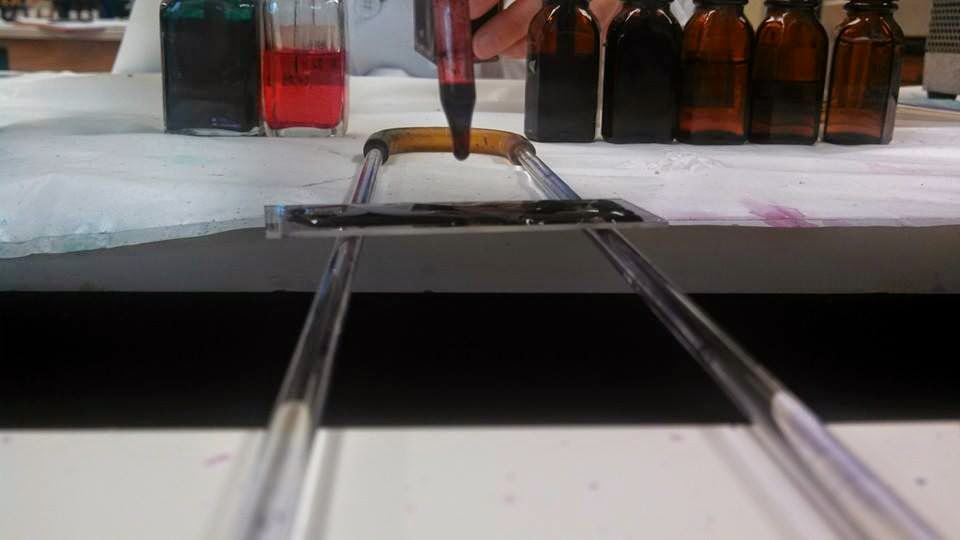
1. Place a small drop of nirgosin stain on one end of the clean microscope slide.
2. Using aseptic technique, transfer bacteria from the agar slant to the drop of nigrosin and mix.

3. Touch the short edge of another microscope slide to the bacteria-nigrosin drop at about a 45 degree angle. Swiftly push the microscope slide across the surface of the first slide, spreading out the drop.
 4. Let the slide air dry.
5. Cover the slide with safranin.
6. Using distilled water, rinse off the excess stain.
7. Blot the slide dry using bibulous paper.
8. Observe the slide under oil immersion microscope.
Results:

Discussion: Our unknown bacteria is not capsulated, because capsule cannot be seen in the negative stain.
No comments:
Post a Comment